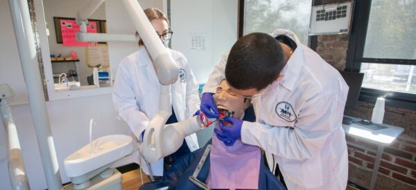
Dental radiology, a critical part of a Dental Assistant’s job responsibilities, is more than just a way to spot a cavity or a chipped tooth. It also helps dentists evaluate bone density, detect issues below the surface, and catch problems that aren’t visible to the naked eye. In some cases, it’s dental radiology that reveals hidden concerns, such as bone loss near the center of a tooth, that would otherwise go undetected.
The International Atomic Energy Agency reports that dental X-rays make up about 26% of all X-rays taken worldwide. Today, we’re looking at two key categories of dental radiology, intraoral (inside) and extraoral (outside), and how they both help support effective diagnosis and treatment.
Intraoral (Inside) Dental Radiology
Intraoral dental radiology refers to X-rays taken from inside the mouth. These are some of the most common and informative types of dental imaging. The main types include:
- Bitewing
- Periapical
- Occlusal View
- Full Mouth Series
These X-rays are taken by placing a film or digital sensor inside the mouth. Patients wear a protective lead apron to minimize radiation exposure (roughly about the same level as you’d get from a cross-country flight). While the dose is small and localized, the information it provides is powerful. Here’s how each type is used:
The Bitewing View
Bitewing X-rays are among the most routine dental images taken. They’re used to evaluate bone levels and detect cavities between the back teeth (molars and premolars). Dentists prefer this view for its accuracy in spotting interproximal decay and early signs of bone loss that can’t be seen with a regular visual exam.
The Periapical View
This view captures the entire tooth, from the visible crown to the tip of the root and surrounding bone. Taken from a distance of about 6–8 inches from the face, it’s especially helpful in diagnosing:
- Cavities
- Gum disease
- Abscesses
- Impacted teeth
- Root or bone fractures
Periapical X-rays are also commonly used in orthodontics to monitor tooth positioning and check for changes after treatment or trauma.
The Intraoral Occlusal View
This type of X-ray provides a broader view of the mouth. It’s taken either from under the chin or angled down from the nose and captures the floor of the mouth or the roof (palate), depending on the angle. The film used is up to four times larger than the size of a standard intraoral film, allowing for a detailed view of:
- Jaw structure
- Cysts or growths
- Tumors
- Misalignments (malocclusions)
While not part of a routine full-mouth series, the occlusal view is helpful for diagnosing more complex or unusual issues.
The Full Mouth Series
This comprehensive set includes up to 18 images, taken during a single visit. It combines both bitewing and periapical views to give a complete picture of a patient’s oral health. It’s typically done for:
- New patients
- Those starting treatment (like implants or major restorations)
- Individuals with a history of dental issues
This series checks not just the teeth, but also nearby tissues and bone, helping dentists spot decay, damage, and other concerns early.
Extraoral (Outside) Radiographs
Extraoral X-rays are taken from outside the mouth and are often used to get a broader view of the teeth, jaw, and facial structure. They’re essential tools in both general dentistry and orthodontics.
Panoramic
Panoramic X-rays capture the entire mouth in a single image. This method was first put into wide use by the U.S. Army to speed up dental screenings. While not as detailed as intraoral views, panoramic images are great for:
- Checking overall oral health
- Spotting impacted teeth
- Assessing jaw alignment
However, they’re less effective for detecting small cavities or pinpointing specific areas of bone loss.
Occlusal Radiographs
These large-view X-rays show the entire arch of teeth in either the upper or lower jaw. They’re used to detect:
- Tooth decay
- Fractures
- Jawbone issues
- Impacted or developing teeth
They can also help evaluate gum health, bite alignment, and identify cysts or tumors.
Cephalometric Radiographs
Also called cephalograms, these side-profile X-rays of the head and neck are essential in orthodontics. They help dentists and orthodontists:
- Analyze jaw and tooth alignment
- Plan for braces or jaw surgery
- Track facial growth in younger patients
- Evaluate how treatments are progressing
They’re especially useful for diagnosing spacing issues, crowding, or jaw discrepancies.
Contact ACI Medical & Dental School Today
While many people think of dental X-rays as a one-size-fits-all tool, there are actually several specialized views, each serving a specific purpose in your oral health. If you’re interested in learning more about dental radiology or starting your certification, contact ACI Medical & Dental School today. We’re here to help you take the next step toward a rewarding career in dental care.


